A Tool Within a Broader Evaluation
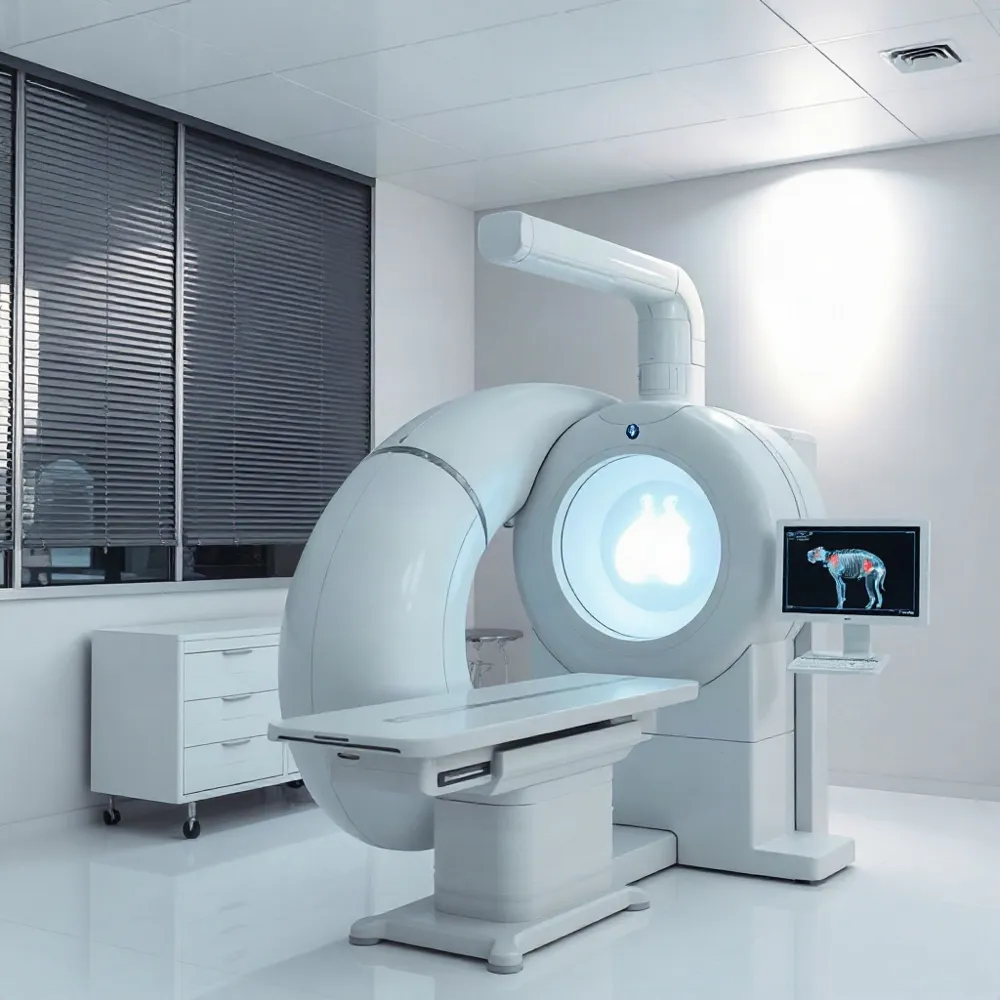
Imaging can help clinicians examine structures that may contribute to pain, such as bones, joints, discs, nerves, and soft tissues. Depending on the suspected cause, a scan may help clarify what to investigate or inform treatment planning.
A scan is only one part of an evaluation. At California Pain Institute, physicians consider imaging alongside a patient’s symptoms, medical history, and physical examination to judge whether a visible finding fits the person’s experience. An abnormality does not automatically explain pain, and a normal scan does not mean the pain is not real.
The right test depends on the clinical question, and imaging is not necessary for everyone with pain. A clinician can weigh whether a scan is likely to help guide decisions and recommend an appropriate next step.
Choosing the Right Imaging Test
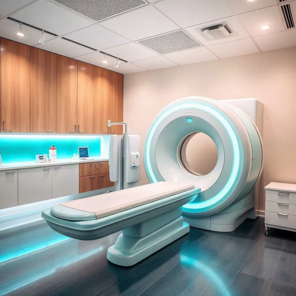 Advanced diagnostic imaging uses specialized technology to create pictures of structures or activity inside the body. Tests may include X-rays, computed tomography (CT), magnetic resonance imaging (MRI), ultrasound, and, in selected situations, nuclear medicine scans such as PET or SPECT. Other studies focus on blood vessels, while fluoroscopy creates moving X-ray images. The choice depends on the clinical question, symptoms, medical history, and examination, rather than a fixed sequence of tests. California Pain Institute describes the role of imaging within its approach to advanced diagnostic techniques.
Each test shows different details. X-rays can reveal bone alignment, fractures, and some degenerative changes. CT creates cross-sectional images and is particularly useful for assessing bone and complex bony anatomy. MRI uses a magnet and radio waves, not ionizing radiation, to show discs, nerves, muscles, ligaments, and other soft tissues. Ultrasound provides real-time views of accessible muscles, tendons, ligaments, joints, and some nerves.
For back pain, a clinician may consider an X-ray, CT, or MRI based on the suspected cause and findings from the evaluation. MRI may be more informative when a disc, nerve, or other soft-tissue problem is suspected. CT uses X-rays and may be preferred for a fracture or complex bone detail, but it involves ionizing radiation. Imaging is not an automatic next step for every episode of back pain; the clinician weighs whether a scan is appropriate and likely to help guide care.
Musculoskeletal ultrasound can assess selected soft-tissue concerns, including tendon or muscle problems, joint fluid, and some nerve issues. Because it shows structures in real time, a clinician may observe them during movement when symptoms are activity-related. Ultrasound can also guide certain injections or other needle procedures. Its usefulness depends on the area and question: it may not show deep structures well and cannot answer every pain-related question.
Reading Results in Clinical Context
A scan shows selected anatomical features, not how intensely a person feels pain or the full experience of the nervous system. Degenerative changes may appear in people who have no symptoms, while significant pain can occur even when imaging looks normal.
Clinicians interpret findings alongside a person’s reported symptoms, medical history, physical examination, and goals. This helps determine whether an abnormality plausibly relates to the pain, rather than treating an incidental finding as its cause. California Pain Institute’s diagnostic approach considers imaging as part of a broader evaluation.
What should I do if my imaging results are normal but I am still in pain?
Normal results do not mean your pain is imaginary. They mean the scan did not show a structural cause it could identify. Arrange follow-up with your clinician and describe where the pain is, what it feels like, what changes it, and how it affects sleep and daily activities.
Routine imaging may not reveal every contributor, including some nerve irritation, functional changes, or nervous-system sensitivity. Depending on the clinical question, further evaluation or treatment may be appropriate. California Pain Institute’s pain specialists assess symptoms and examination findings alongside imaging when considering next steps.
Seek urgent care for severe or rapidly worsening symptoms, new weakness, loss of bladder or bowel control, or other sudden concerning changes.
When Imaging May Be Appropriate
 A scan is not usually the first step for new or uncomplicated low-back pain, even when discomfort travels down a leg. Without warning signs, many cases improve with conservative care, and imaging may not change the initial approach. The approach to evaluating acute lower back pain explains why a clinician may recommend observation and noninvasive treatment before ordering a scan.
A clinician may consider imaging sooner if symptoms or examination findings raise concern for a serious condition, such as a fracture, infection, cancer, or compression of nerves in the lower spine. New or worsening numbness, weakness, or other neurological problems, a significant injury, or pain that continues to worsen despite appropriate treatment can also affect the decision.
If pain persists or worsens after about six weeks of appropriate care, imaging may be considered when a procedure or surgery is being discussed. MRI, typically without contrast, can help assess structures relevant to that decision. At California Pain Institute, evaluation includes the patient’s history and physical examination alongside imaging when it may clarify the suspected cause and inform care.
Some symptoms require urgent medical assessment rather than a routine appointment. Seek immediate care for new loss of bladder or bowel control, numbness around the groin or saddle area, or rapidly worsening leg weakness, as these may signal serious nerve compression.
The decision should be individualized: scans can reveal common structural changes that are not causing a person’s pain, sometimes prompting unnecessary follow-up. A pain specialist can weigh symptoms, examination findings, and how imaging results could affect treatment before recommending a test.
Understanding Persistent and Chronic Pain
Pain is generally considered chronic when it persists or recurs for longer than three months. Pain lasting one to three months is often called subacute, although terminology can vary by condition. The three-month mark is a general guide, not a diagnosis of the cause or severity. For more on how chronic pain is described and assessed, see understanding chronic pain and its impact.
Pain can continue after an injury has healed, arise from several contributing factors, or persist without a clearly identified cause. A scan may show a structural change, but that finding alone does not explain how or why someone hurts. At California Pain Institute, a pain evaluation considers symptoms and medical history alongside examination and any relevant imaging.
How long does pain need to last to be considered chronic?
The duration is only one part of the picture. If pain persists, returns repeatedly, or interferes with daily activities, discuss it with a healthcare professional. A clinician can assess possible causes and decide whether further evaluation, including imaging, could help guide care.
What are common types and causes of chronic pain conditions?
Common forms include back or neck pain, arthritis and other joint pain, headaches or migraines, nerve pain, fibromyalgia, and cancer-related pain. Symptoms may begin after an injury, surgery, or infection, or occur with an underlying condition such as arthritis or spinal stenosis. Sometimes pain continues after the original cause has healed, or no single cause can be identified.
Changes in how the nervous system processes pain can also contribute. Poor sleep, stress, anxiety, or depression may accompany chronic pain and affect a person’s experience, but they do not make the pain less real. California Pain Institute’s assessment-led approach takes the person’s symptoms and circumstances into account rather than treating an imaging result as the whole explanation.
Turning Evaluation Into a Care Plan
 What treatment options are available for chronic pain? A care plan should reflect the person’s symptoms, medical history, daily needs, and goals. Imaging may clarify relevant anatomy or support a suspected diagnosis, but a scan alone does not determine treatment. At California Pain Institute, pain specialists use clinical evaluation, including imaging when appropriate, to inform diagnosis and care planning.
Depending on the cause, care may include treating an underlying condition, medication when appropriate, physical therapy, and exercise tailored to the patient’s abilities. Sleep strategies and gradually increasing activity may also support daily function. Cognitive behavioral therapy can help people build coping skills and address pain’s effects on mood and everyday life.
Selected image-guided procedures, such as injections or nerve blocks, may be considered when they fit the suspected cause and pattern of pain. Imaging can help guide placement, but whether a procedure is suitable depends on the full clinical picture. California Pain Institute describes the use of imaging guidance as part of focused pain diagnosis and targeted procedures, rather than as a substitute for individualized assessment.
Follow-up imaging may be appropriate to assess healing, evaluate an intervention, or monitor a change that could affect care. It is not a routine requirement for everyone. A coordinated plan can be reviewed over time and adjusted according to symptoms, function, treatment response, and the patient’s priorities.
Imaging Supports Individualized Care
Imaging is most useful when it addresses a focused clinical question. A pain specialist at capaininstitute.com considers scan findings alongside a person’s symptoms, medical history, and examination, rather than treating an image as a diagnosis on its own.
Before a scan, ask your healthcare professional what it may clarify and how the result could change your care. This discussion can help you weigh the potential value of imaging and understand how it fits into an individualized evaluation.